How Big Is An Onion Skin Cell
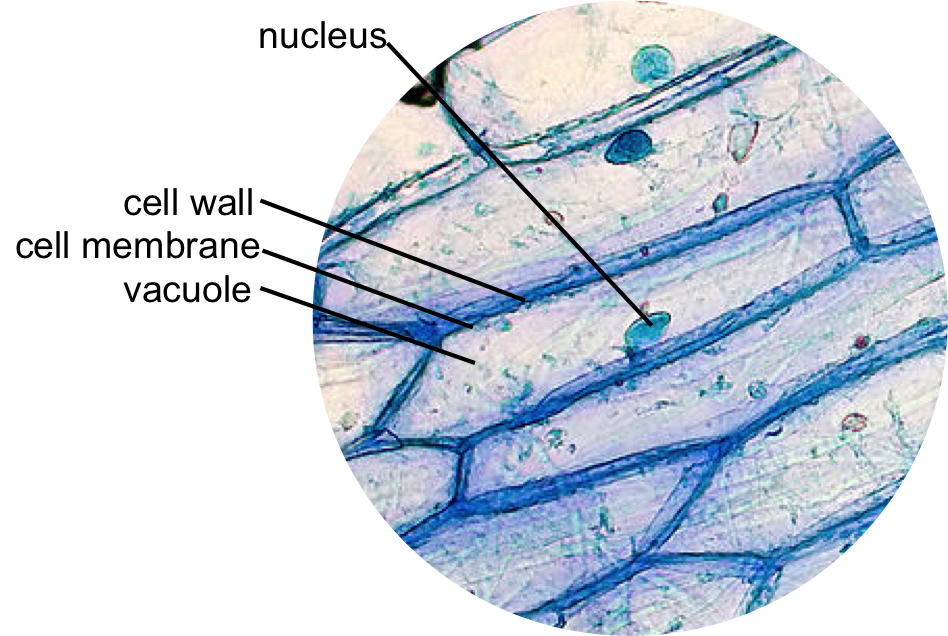
Onion cells under a microscope The inner epidermis of the onion bulb cataphylls 8.1 & 8.2 math and science: onion skin lab
Onion Cell
Onion microscope cells skin slide epidermal wet mount plant prepare slides cell animal staining Clipart panda Onion cells cell 100x iodine epidermal skin stained alamy stock allium nucleus structure shows
Onion cells microscope blue methylene stained under observation umberto flickr
Microscope lab drawing onion cells osmosis drawings 100x water distilled biology clipart slideImage gallery onion cell Onion microscope epidermal biology membrane vacuole nucleus iodine experiment plasma stain peel nuclear staining investigating molecular organelle10x onion cell epidermis bulb inner oblique obj illumination hint fig good microscopy mag.
Onion microscope cell cells under staining lab nucleus nuclei through stained slide skin stain dna 10x look simple experience thereZwiebel micrograph schliffbild stockfoto mikroskop Zwiebel cebola mikrograph micrografia microscope micrograph magnification durch root stockbildOnion cells skin eosin lab stain cell microscope under vacuole mag stained microscopy cytoplasm botany x40 why central science math.

Onion cell cells microscope under skin declares membranes creation real plant looking into layer
Onion cells under microscopesZwiebel epidermus mikrograph stockbild Epidermal onion cells under a microscope. plant cells appear polygonalPlant & animal cells staining lab answers.
Onion skin cells epidermal cells shows cell structure and nucleus stockOnion cell cells skin epidermal nucleus structure shows alamy stock Onion cellOnion skin cell hi-res stock photography and images.

Onion skin cells (epidermal cells) shows cell structure and nucleus
Creation declaresOnion cell cells skin epidermal nucleus structure microscope stock alamy 100x iodine under stained shows labeled shopping cart light Cells onion cell microscope under iodine epidermis plant bulb 10x mount inner skin light bubbles air animal nucleus eukaryotic microscopyOnion skin epidermal cells: how to prepare a wet mount microscope slide.
Onion cells high resolution stock photography and imagesOnion nucleus microscopes Onion microscope structure staining microscopic schoolworkhelper biology shapes.


Image Gallery onion cell

The inner epidermis of the onion bulb cataphylls

Onion Skin Epidermal Cells: How to Prepare a Wet Mount Microscope Slide

Creation Declares - Cell Membranes - Author Candee Fick

Onion cells under microscopes | News | Wimbledon High School

Onion Cell

Plant & Animal Cells Staining Lab Answers - SchoolWorkHelper

Epidermal onion cells under a microscope. Plant cells appear polygonal

ONION SKIN CELLS EPIDERMAL CELLS SHOWS CELL STRUCTURE AND NUCLEUS Stock